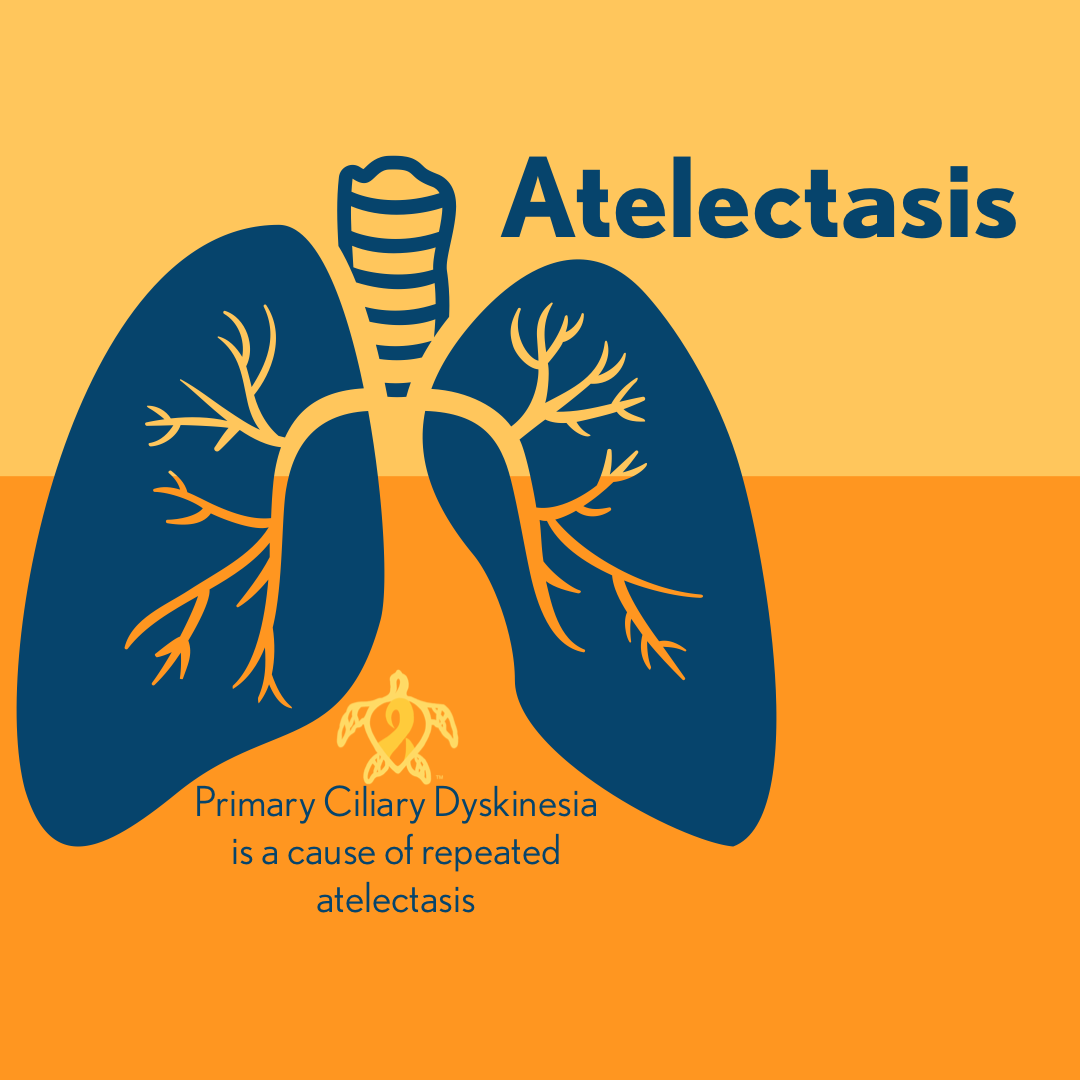
Atelectasis is a complete or partial collapse of the lung, a lung lobe, or partial lung lobe (segment). In atelectasis the lung or lung segment does not inflate and the air sacs or alveoli are no longer filled with air. This section of the lungs can not take part in gas exchange any longer. If the atelectasis comes on gradually and is relatively a small collapse, then the other areas of the lungs can take up slack for the collapsed area and adequately supply the body with the required gas exchange needed. Rapid onset of atelectasis, especially if the area of collapse is big, can lead to shortness of breath, rapid heart rate, coughing, and cyanosis. There usually isn’t pain associated with atelectasis unless the area of atelectasis is big; or the pain is associated with the cause of the atelectasis like pneumonia, injury, or even the obstruction causing the atelectasis itself. Children with atelectasis may appear agitated, anxious, or even scared. Complications of atelectasis can include low blood oxygen, pneumonia, and respiratory failure. The symptoms of atelectasis may reflect the underlying condition that initially caused the atelectasis. Underlying medical conditions that are often associated with atelectasis are conditions that cause respiratory weakness, primary ciliary dyskinesia, cystic fibrosis, lung tumor, lung cancer, conditions that cause fluid build up in the lungs, lung scar tissue, airway blockage, chest injuries, and atelectasis can even be a complication after a surgical procedure.
Atelectasis has many causes and typically occurs when the lungs can not expand and fill with air. Atelectasis is very common after surgery where the patient is given medication to sleep and stifle their cough responses. Limiting a patient’s ability to cough allows mucus to fill and sit in the airways which can lead to mucus plugging and or pneumonia. Those areas of infection and or mucus plugging can become obstructions which trigger an episode of atelectasis. Airway blockages are another cause of atelectasis. Blockages can be caused any number of things like a mucus plug, foreign objects (popcorn), cancer, poorly place ventilator tubing, and many other things. Once the airway has been blocked off, and the air in the air sacs have already been exchanged into the body new air cant get past the blockage, so the air sacs deflate. Factors that increase a patient’s risks of atelectasis is age, having a condition that interferes with spontaneous coughing, confinement to bed, having a condition that impairs swallowing function, lung diseases (like cystic fibrosis, primary ciliary dyskinesia, bronchiectasis, COPD, and asthma), premature birth, recent abdominal or chest surgery, recent general anesthesia, having a condition that causes weak respiratory muscles (like muscular dystrophy, SMA, spinal cord injury, or so on), and having any condition that can cause shallow breathing (like medication side effects or mechanical limitations like shallow breathing due to a rib fracture.
The main goals of treatment in atelectasis are to treat the cause of the condition and to reinflate the lung. Treatments will vary based on the underlying condition that caused the atelectasis in the first place. Airway obstructions can be cleared with a surgical procedure known as a bronchoscopy, usually reserved for foreign objects, or mucus plugging that does not clear up with manual chest percussion and autogenic drainage. For the most part atelectasis is usually treated with deep breathing exercises, coughing, autogenic drainage techniques, chest percussion, and breathing medications. If the atelectasis is caused by chest pressure, the doctors will remove the cause of the pressure; usually a tumor or fluid build up in the chest cavity. In cases where those things are not enough surgery (bronchoscopy) or medical devices such as PEEP (positive end-expiratory pressure) or a CPAP (continuous positive airway pressure) may be prescribed.
Atelectasis appears to be a common condition associated with PCD (primary ciliary dyskinesia). This is surmised due to the ineffective mucus clearance that affects PCD patients. Mucus in PCD that can not be cleared becomes thick and oftentimes leads to plugging. The ineffective mucus clearance in PCD can also lead to pneumonia and other types of respiratory infections that can lead to obstructions in the lungs and trigger an episode of atelectasis. Atelectasis can be periodic and last for a short or a long amount of time; including several years. In PCD areas of the lungs most effected by atelectasis are the right middle lobe, left lingula, and both the right and lower lobes. However; lobar or segmental atelectasis with associated bronchiectasis in the middle lobes is more common in childhood PCD.
Be sure to visit us next week for another Topic Thursday!
Join our Facebook group Turtle Talk Café today, click here.
We have several ways that you can donate to PCD Smiles;
- Visit Smile E. Turtle's Amazon Wishlist
- For more information on how you can donate, please visit our "Donation" page to check out our "Do & Don't policies.
- Or sponsor a PCD Smiles cheer package today!
- To shop for your “Official” turtle care ribbon gear today, visit PCD Style or Smile E. Cove
Thank you for your consideration!


